The transillumination possibility of imidazole–osmium postfixed tissue and its consequences for the handling of tissue samples
- Voigt, Tilman Anatomy Unit, Department of Medicine, University of Fribourg, Switzerland
- Dauber, Wolfgang Institute of Anatomy, Eberhard-Karls-University Tübingen, Germany
-
28.01.2005
Published in:
- Microscopy and Microanalysis. - 2005, vol. 11, no. 1, p. 42-45
English
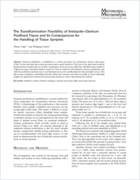
Osmium postfixation is established as a routine procedure for transmission electron microscopy (TEM). On the one hand, this routine procedure leads to good results for TEM, but on the other hand results in blackened tissue samples that do not allow examination of any structures within the embedded tissue sample by a light microscope. Equivalent fixation results for TEM are achieved with imidazole–osmium postfixation, and with this postfixation method tissue is not blackened and can be transilluminated with point light sources. This allows easier recognition of histological details within tissue samples and makes it possible to screen embedded samples for appropriate ultrastructural processing. Jejunum is used to demonstrate the method.
- Faculty
- Faculté des sciences et de médecine
- Department
- Département de Médecine
- Language
-
- English
- Classification
- Biological sciences
- License
-
License undefined
- Identifiers
-
- RERO DOC 5168
- DOI 10.1017/S1431927605050117
- Persistent URL
- https://folia.unifr.ch/unifr/documents/299988
Statistics
Document views: 163
File downloads:
- Texte intégral: 260