3D imaging of undissected optically cleared Anopheles stephensi mosquitoes and midguts infected with Plasmodium parasites
- Niz, Mariana De Institute of Cell Biology, Heussler Research Group, University of Bern, Bern, Switzerland
- Kehrer, Jessica Center for Infectious Diseases, Integrative Parasitology, Heidelberg University Medical School, Heidelberg, Germany,
- Brancucci, Nicolas M. B. Wellcome Centre for Integrative Parasitology, Institute of Infection, Immunity & Inflammation, University of Glasgow, Glasgow, United Kingdom
- Moalli, Federica Theodor Kocher Institute, University of Bern, Bern, Switzerland
- Reynaud, Emmanuel G. School of Biomolecular and Biomedical Science, University College Dublin, Dublin, Ireland
- Stein, Jens V. Department of Oncology, Microbiology and Immunology, University of Fribourg, Fribourg, Switzerland
- Frischknecht, Friedrich Center for Infectious Diseases, Integrative Parasitology, Heidelberg University Medical School, Heidelberg, Germany,
- 16.09.2020
Published in:
- PLOS ONE. - 2020, vol. 15, no. 9, p. e0238134
English
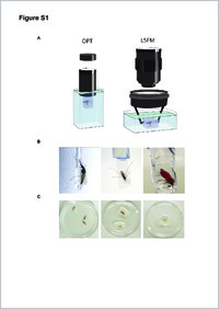
Malaria is a life-threatening disease, caused by Apicomplexan parasites of the Plasmodium genus. The Anopheles mosquito is necessary for the sexual replication of these parasites and for their transmission to vertebrate hosts, including humans. Imaging of the parasite within the insect vector has been attempted using multiple microscopy methods, most of which are hampered by the presence of the light scattering opaque cuticle of the mosquito. So far, most imaging of the Plasmodium mosquito stages depended on either sectioning or surgical dissection of important anatomical sites, such as the midgut and the salivary glands. Optical projection tomography (OPT) and light sheet fluorescence microscopy (LSFM) enable imaging fields of view in the centimeter scale whilst providing micrometer resolution. In this paper, we compare different optical clearing protocols and present reconstructions of the whole body of Plasmodium-infected, optically cleared Anopheles stephensi mosquitoes and their midguts. The 3D-reconstructions from OPT imaging show detailed features of the mosquito anatomy and enable overall localization of parasites in midguts. Additionally, LSFM imaging of mosquito midguts shows detailed distribution of oocysts in extracted midguts. This work was submitted as a pre-print to bioRxiv, available at https://www.biorxiv.org/content/10.1101/682054v2.
- Faculty
- Faculté des sciences et de médecine
- Department
- Département de Médecine
- Language
-
- English
- Classification
- Biological sciences
- License
-
License undefined
- Identifiers
-
- RERO DOC 329643
- DOI 10.1371/journal.pone.0238134
- Persistent URL
- https://folia.unifr.ch/unifr/documents/309097
Other files
Statistics
Document views: 153
File downloads:
- pdf: 288
- Supplementary material: 162