Zebrafish fin regeneration after cryoinjury-induced tissue damage
- Chassot, Bérénice Department of Biology, University of Fribourg, Switzerland
- Pury, David Department of Biology, University of Fribourg, Switzerland
- Jaźwińska, Anna Department of Biology, University of Fribourg, Switzerland
-
15.06.2016
Published in:
- Biology Open. - 2016, vol. 5, no. 6, p. 819–828
English
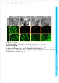
Although fin regeneration following an amputation procedure has been well characterized, little is known about the impact of prolonged tissue damage on the execution of the regenerative programme in the zebrafish appendages. To induce histolytic processes in the caudal fin, we developed a new cryolesion model that combines the detrimental effects of freezing/thawing and ischemia. In contrast to the common transection model, the damaged part of the fin was spontaneously shed within two days after cryoinjury. The remaining stump contained a distorted margin with a mixture of dead material and healthy cells that concomitantly induced two opposing processes of tissue debris degradation and cellular proliferation, respectively. Between two and seven days after cryoinjury, this reparative/proliferative phase was morphologically featured by displaced fragments of broken bones. A blastemal marker msxB was induced in the intact mesenchyme below the damaged stump margin. Live imaging of epithelial and osteoblastic transgenic reporter lines revealed that the tissue- specific regenerative programmes were initiated after the clearance of damaged material. Despite histolytic perturbation during the first week after cryoinjury, the fin regeneration resumed and was completed without further alteration in comparison to the simple amputation model. This model reveals the powerful ability of the zebrafish to restore the original appendage architecture after the extended histolysis of the stump.
- Faculty
- Faculté des sciences et de médecine
- Department
- Département de Biologie
- Language
-
- English
- Classification
- Biological sciences
- License
-
License undefined
- Identifiers
-
- RERO DOC 277316
- DOI 10.1242/bio.016865
- Persistent URL
- https://folia.unifr.ch/unifr/documents/305230
Other files
Statistics
Document views: 133
File downloads:
- pdf: 330
- Supplementary material: 166