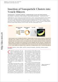
Insertion of Nanoparticle Clusters into Vesicle Bilayers
- Bonnaud, Cécile Adolphe Merkle Institute, University of Fribourg, Switzerland - Chemistry Department, University of Fribourg, Switzerland
- Monnier, Christophe A. Adolphe Merkle Institute, University of Fribourg, Switzerland
- Demurtas, Davide Interdisciplinary Center for Electron Microscopy, Ecole Polytechnique Fédérale de Lausanne, Switzerland
- Jud, Corinne Adolphe Merkle Institute, University of Fribourg, Switzerland
- Vanhecke, Dimitri Adolphe Merkle Institute, University of Fribourg, Switzerland
- Montet, Xavier Radiology Department, Geneva University Hospital, Rue Gabrielle-Perret-Gentil 4, 1211 Genève 14, Switzerland
- Hovius, Ruud Laboratory of Physical Chemistry of Polymers and Membranes, Ecole Polytechnique Fédérale de Lausanne, Station 6, 1015 Lausanne, Switzerland
- Lattuada, Marco Adolphe Merkle Institute, University of Fribourg, Switzerland
- Rothen-Rutishauser, Barbara Adolphe Merkle Institute, University of Fribourg, Switzerland - Respiratory Medicine, Bern University Hospital, Inselspital, Freiburgstrasse, 3010 Bern, Switzerland
- Petri-Fink, Alke Adolphe Merkle Institute, University of Fribourg, Switzerland - Chemistry Department, University of Fribourg, Switzerland
-
22.04.2014
Published in:
- ACS Nano. - 2014, vol. 8, no. 4, p. 3451–3460
Liposomes
Vesicles
SPIONs
Cryo TEM
Cryo electron tomography
Self assembly
Membrane energetics
Drug delivery
English
A major contemporary concern in developing effective liposome–nanoparticle hybrids is the present inclusion size limitation of nanoparticles between vesicle bilayers, which is considered to be around 6.5 nm in diameter. In this article, we present experimental observations backed by theoretical considerations which show that greater structures can be incorporated within vesicle membranes by promoting the clustering of nanoparticles before liposome formation. Cryo-transmission electron microscopy and cryo-electron tomography confirm these observations at unprecedented detail and underpin that the liposome membranes can accommodate flexible structures of up to 60 nm in size. These results imply that this material is more versatile in terms of inclusion capabilities and consequently widens the opportunities in developing multivalent vesicles for nanobiotechnology applications.
- Faculty
- Faculté des sciences et de médecine
- Department
- Département de Chimie
- Language
-
- English
- Classification
- Physics
- License
-
License undefined
- Identifiers
-
- RERO DOC 210017
- DOI 10.1021/nn406349z
- Persistent URL
- https://folia.unifr.ch/unifr/documents/303470
Other files
Export to…
Statistics
Document views: 250
File downloads:
- pdf: 430
- Supplementary material: 241