Atomic-resolution structure of a disease-relevant Aβ(1-42) amyloid fibril.
- Wälti MA Laboratorium für Physikalische Chemie, Eidgenössische Technische Hochschule Zürich, 8093 Zurich, Switzerland;
- Ravotti F Laboratorium für Physikalische Chemie, Eidgenössische Technische Hochschule Zürich, 8093 Zurich, Switzerland;
- Arai H Department of Molecular Biology and Biochemistry, University of California, Irvine, CA 92697;
- Glabe CG Department of Molecular Biology and Biochemistry, University of California, Irvine, CA 92697; Biochemistry Department, Faculty of Science and Experimental Biochemistry Unit, King Fahd Medical Research Center, King Abdulaziz University, Jeddah 21589, Saudi Arabia;
- Wall JS Brookhaven National Laboratory, Upton, NY 11973-5000;
- Böckmann A Institut de Biologie et Chimie des Protéines, Bases Moléculaires et Structurales des Systèmes Infectieux, Labex Ecofect, UMR 5086 CNRS, Université de Lyon, 69007 Lyon, France; a.bockmann@ibcp.fr beme@ethz.ch roland.riek@phys.chem.ethz.ch.
- Güntert P Laboratorium für Physikalische Chemie, Eidgenössische Technische Hochschule Zürich, 8093 Zurich, Switzerland; Institute of Biophysical Chemistry, Center for Biomolecular Magnetic Resonance, Goethe University Frankfurt am Main, 60438 Frankfurt am Main, Germany; Department of Chemistry, Graduate School of Science and Engineering, Tokyo Metropolitan University, Hachioji, Tokyo 192-0397, Japan.
- Meier BH Laboratorium für Physikalische Chemie, Eidgenössische Technische Hochschule Zürich, 8093 Zurich, Switzerland; a.bockmann@ibcp.fr beme@ethz.ch roland.riek@phys.chem.ethz.ch.
- Riek R Laboratorium für Physikalische Chemie, Eidgenössische Technische Hochschule Zürich, 8093 Zurich, Switzerland; a.bockmann@ibcp.fr beme@ethz.ch roland.riek@phys.chem.ethz.ch.
- 2016-07-30
Published in:
- Proceedings of the National Academy of Sciences of the United States of America. - 2016
Alzheimer’s disease
amyloid
protein structure
solid-state NMR
Amyloid beta-Peptides
Cloning, Molecular
Escherichia coli
Gene Expression
Genetic Vectors
Humans
Microscopy, Electron
Models, Molecular
Nuclear Magnetic Resonance, Biomolecular
Peptide Fragments
Protein Conformation, beta-Strand
Recombinant Proteins
English
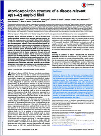
Amyloid-β (Aβ) is present in humans as a 39- to 42-amino acid residue metabolic product of the amyloid precursor protein. Although the two predominant forms, Aβ(1-40) and Aβ(1-42), differ in only two residues, they display different biophysical, biological, and clinical behavior. Aβ(1-42) is the more neurotoxic species, aggregates much faster, and dominates in senile plaque of Alzheimer's disease (AD) patients. Although small Aβ oligomers are believed to be the neurotoxic species, Aβ amyloid fibrils are, because of their presence in plaques, a pathological hallmark of AD and appear to play an important role in disease progression through cell-to-cell transmissibility. Here, we solved the 3D structure of a disease-relevant Aβ(1-42) fibril polymorph, combining data from solid-state NMR spectroscopy and mass-per-length measurements from EM. The 3D structure is composed of two molecules per fibril layer, with residues 15-42 forming a double-horseshoe-like cross-β-sheet entity with maximally buried hydrophobic side chains. Residues 1-14 are partially ordered and in a β-strand conformation, but do not display unambiguous distance restraints to the remainder of the core structure.
- Language
-
- English
- Open access status
- bronze
- Identifiers
-
- DOI 10.1073/pnas.1600749113
- PMID 27469165
- Persistent URL
- https://folia.unifr.ch/global/documents/181116
Statistics
Document views: 16
File downloads:
- fulltext.pdf: 0